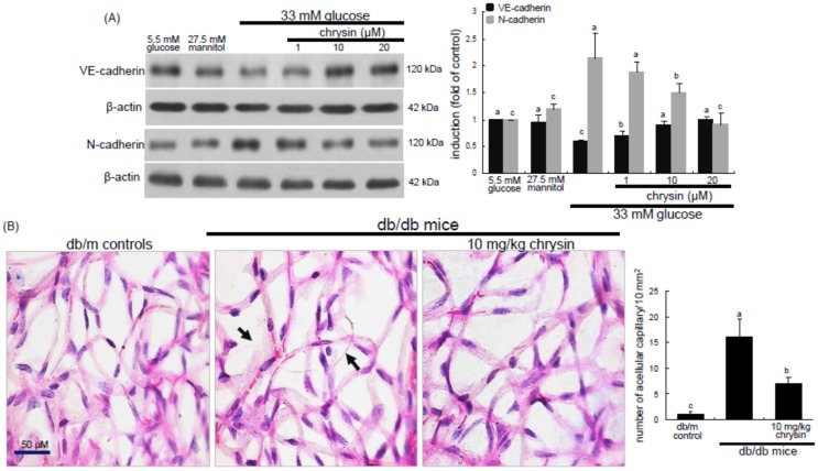
Figure 5.
Effect of chrysin on expression of VE-cadherin and N-cadherin in high glucose-exposed HRMVEC (A) and histopathological changes in trypsin-digested retinal vessels (B). HRMVEC were incubated with 33 mM glucose in the absence and presence of 1–20 μM chrysin up to five days. Cells were also incubated with 5.5 mM glucose and 27.5 mM mannitol as osmotic controls. Cell lysates were subject to Western blot analysis using a primary antibody against VE-cadherin or N-cadherin (A). β-actin protein was used as an internal control. Bar graphs (mean ± SEM, n = 3) in the right panels represent densitometric results of left blot bands. The db/db mice were orally treated with 10 mg/kg chrysin for 10 weeks, and db/m mice were introduced as control animals. Retinal vessels were stained with H&E stain (B). Acellular capillaries (black arrow) were observed in db/db mice. Magnification: 200×. The number of acellular capillaries was measured to assess the extent of retinopathy. Values in bar graphs (mean ± SEM, n = 3) not sharing a common letter differ, p < 0.05.

