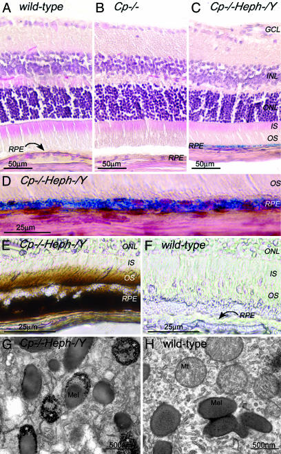
Fig. 2.
Adult (6-month-old) Cp–/–Heph–/Y RPE and photoreceptors accumulate iron. (A–C) 6-month-old WT (A), Cp–/– (B), and Cp–/–Heph–/Y (C) retinas Perls' stained for iron (blue) and counterstained with hematoxylin/eosin. (D) High magnification of Prussian blue Perls' label in 6-month-old Cp–/–Heph–/Y RPE. (E and F) Light photomicrographs of 6-month-old Cp–/–Heph–/Y (E) and WT (F) retinas after DAB enhancement (brown) of Perls' stain. (G–H) Electron micrographs of RPE from 6-month-old Cp–/–Heph–/Y (G) and WT (H) eyes. Only the Cp–/–Heph–/Y RPE (G) contains electrondense vesicles (*) sometimes fused with melanosomes.

