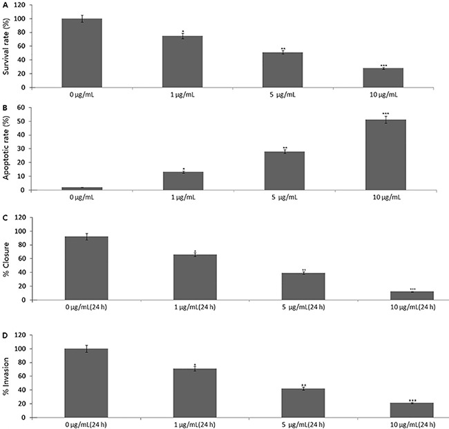
Figure 1. Results of ES-2 cells treated with 1, 5, and 10 μg/mL propofol for 24 h. A, Cell viability by MTT assay. B, Cell apoptosis using Annexin V-staining followed by a FACScan flow cytometer assay. C, Migration by wound healing migration assay. D, Transwell invasion assay vs untreated cells (0 μg/mL). *P<0.05, **P<0.01, ***P<0.001 (t-test).

