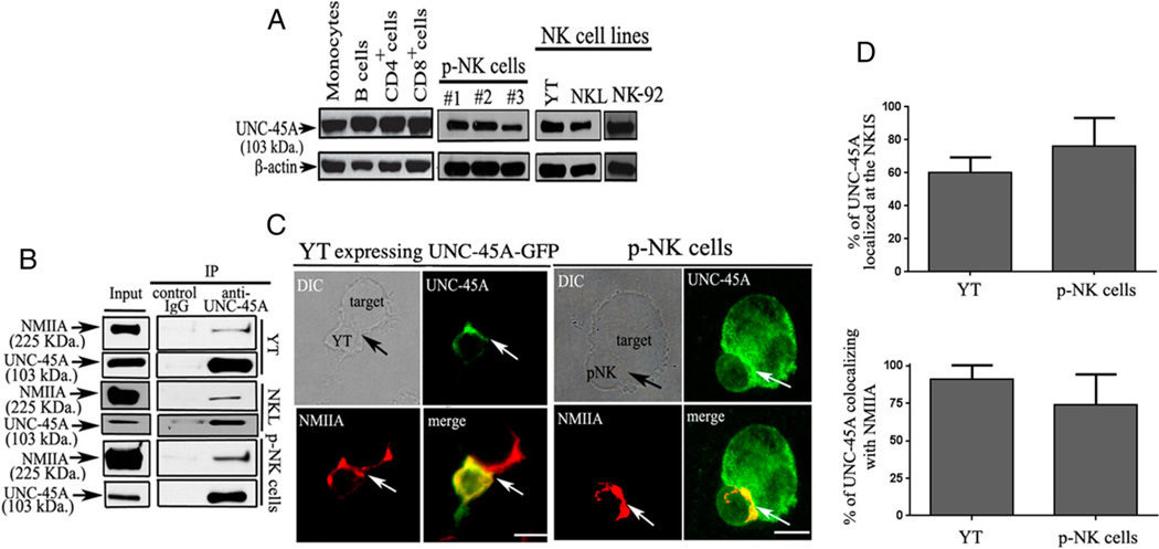
FIGURE 1.
UNC-45A expression and localization in NK cells. (A) Western blot analysis of UNC-45A in lysates from primary monocytes, CD19+ B cells, CD4+ T cells, CD8+ T cells, and CD3C−D56+ NK cells from the peripheral blood and the YT, NKL, and NK-92 NK cell lines. β-Actin was used as loading control. The blots presented are representative of three independent experiments with three different blood donors. (B) UNC-45A immunoprecipitated from lysates of YT and NKL NK cell lines or primary p-NK cells with an anti-UNC-45A mAb. Coimmunoprecipitated NMIIA was detected by Western blot analysis using an anti-NMIIA polyclonal Ab. Immunoprecipitation with nonspecific IgG was performed as a control. (C, left panels) YT NK cells overexpressing a UNC-45A–GFP fusion protein were incubated with 721.221 target cells. After fixation, cells were stained with an anti-NMIIA polyclonal Ab and Texas red goat anti-rabbit IgG and analyzed by fluorescence microscopy. (Right panels) p-NK cells incubated with K562 target cells. After fixation, cell conjugates were stained with anti–UNC-45A and anti-NMIIA Abs followed by FITC-conjugated donkey anti-mouse IgG or by Texas red-conjugated goat anti-rabbit IgG for UNC-45A and NMIIA, respectively, and analyzed by fluorescence microscopy. Arrows indicate NKISs. Scale bars, 5 µm. (D, top panel) Quantification of the percentage of UNC-45A localized at the NKIS out of the total present in the cells for YT and p-NK. (Bottom panel) Quantification of the percentage of UNC-45A colocalizing with NMIIA out of the total present in the cells for YT and p-NK.

