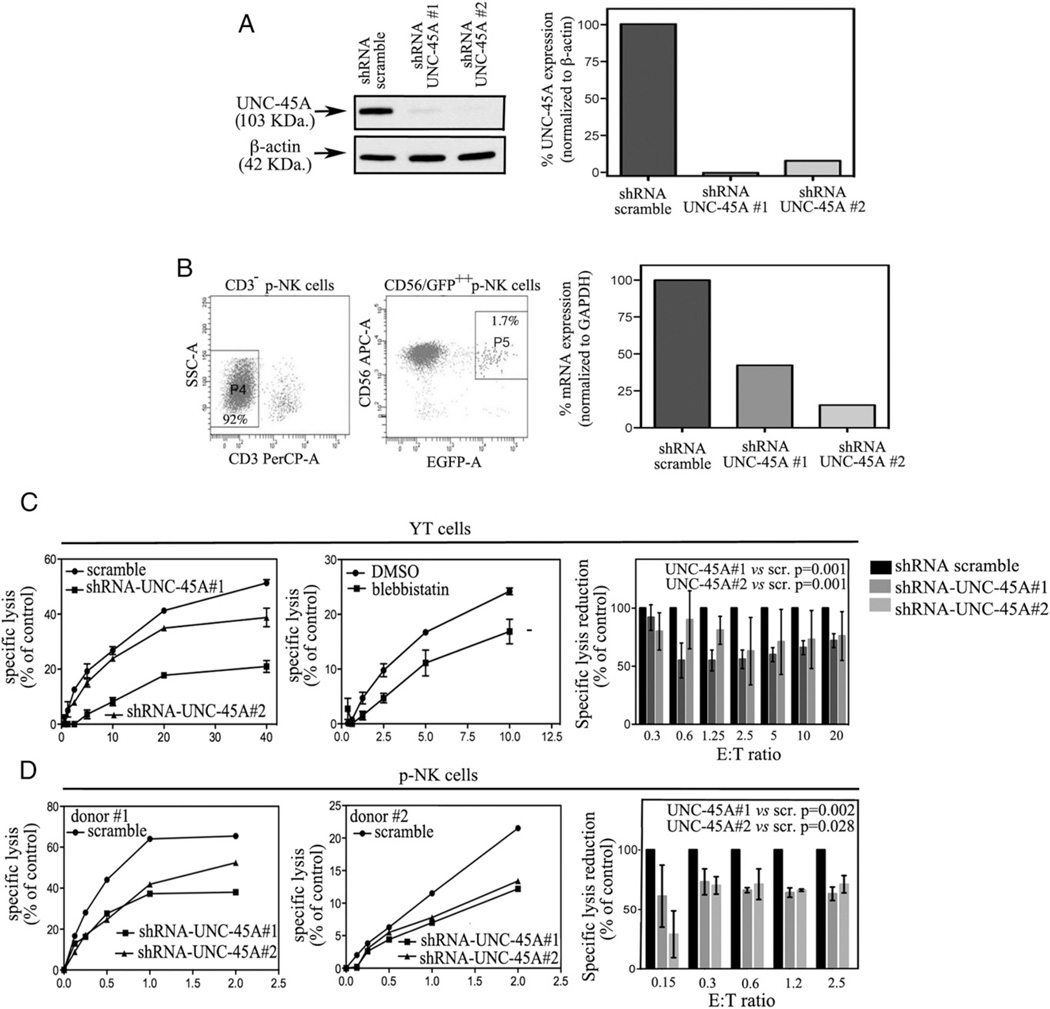
FIGURE 3.
UNC-45A knockdown hinders NK cell–mediated cytotoxicity. (A, left panel) Western blot analysis of UNC-45A expression in YT cells transduced with either shRNA-scramble or shRNA targeting two different UNC-45A regions (1 and 2). β-Actin was used as a loading control. (Right panel) Quantification of the percentage of UNC-45A expression normalized to β-actin. (B, left panels) UNC-45A knockdown in primary CD3− lymphocytes (P4) virally infected with lenti-pEF-UNC45-shRNAmir (1 and 2). Forty-eight hours postinfection, GFP+ CD3−CD56+ cells were sorted (P5) for quantification of UNC-45A levels. (Right panel) Efficiency of UNC-45A knockdown in primary NK cells measured by quantitative RT-PCR. The mRNA values reported are relative to the control gene (GAPDH) and normalized to control-treated cells. (C, left panel) Residual specific lysis in YT cells infected with lentiviral particles carrying either shRNA scramble or shRNAs directed against two different UNC-45A sequences (1 and 2) incubated with 721.221 target cells at the indicated ratios. A representative experiment is shown. (Middle panel) Residual specific lysis in YT cells either mock- (DMSO) or blebbistatin (75 µM)-treated and incubated with 721.221 target cells at the indicated ratios. A representative experiment is shown. (Right panel) Average of five independent experiments reported as reduction in specific lysis as compared with the controls (set at 100%). (D, left and middle panels) Residual specific lysis in lenti-pEF-UNC45-shRNAmir (1 and 2) or scramble infected, CD3−/CD56+/GFP+-sorted p-NK cells derived from healthy donors (donors 1 and 2) incubated with K562 target cells at the indicated ratios. (Right panel) Average of three independent experiments reported as reduction compared with controls (set at 100%). For all experiments (conducted in triplicates), specific lysis was assessed by [51CR]-release assay. Statistical significance was set at p = 0.05.

