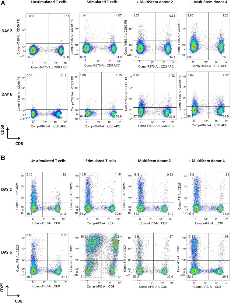
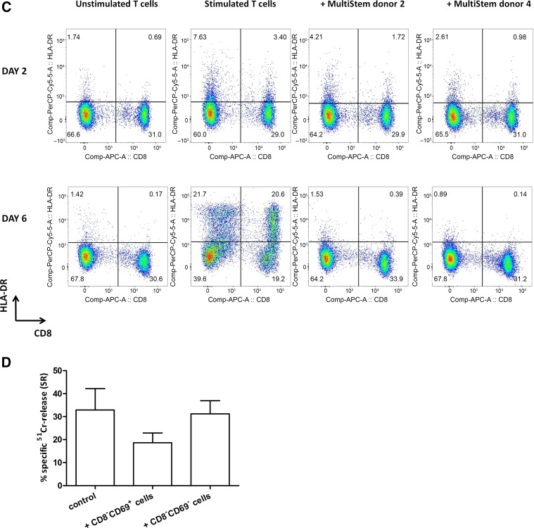
Figure 6.
T-cell activation marker expression is altered in the presence of MultiStem. (A–C): Flow-cytometric analysis of CD3+ T lymphocytes for expression of T-cell activation markers CD69 (A), CD25 (B), and HLA-DR (C) on days 2 and 6 of a 6-day stimulation period with irradiated (40 Gy) allogeneic Epstein-Barr virus-positive (EBV+) B cells (stimulator:responder [S:R] ratio of 1:20) in the absence [(un)stimulated T cells] or presence (+MultiStem donor 2/4) of irradiated (30 Gy) third-party MultiStem cells (suppressor:responder ratio of 1:1). Data are presented as percentage of cells within the CD3+ lymphocyte gate. One representative experiment with one T-cell donor and two different MultiStem donors (donors 2 and 4) out of three independent experiments is shown. (D): Freshly isolated CD3+ T cells were primed with irradiated (40 Gy) allogeneic EBV+ B cells (S:R ratio of 1:20) in the presence of irradiated (30 Gy) third-party MultiStem cells (ratio of 1:2). After 6 days of stimulation, two populations were sorted from the coculture (CD45+CD3+CD8−CD69+ T cells and CD45+CD3+CD8−CD69− T cells). These fractions were added at a ratio of 1:2 to a subsequent mixed-lymphocyte culture with freshly isolated responder T cells and irradiated allogeneic EBV+ B cells (S:R ratio of 1:20). After 7 days, anti-CD3-redirected cytotoxicity was analyzed. Data are expressed as mean ± SEM percentage of anti-CD3-dependent specific 51Cr-release (% SR) of P815 target cells (effector:target ratio 10:1). Results are pooled from two independent experiments, in which two different T-cell donors and two MultiStem donors (donors 2 and 3) were used. Abbreviations: APC, allophycocyanin; Comp, compensation; cy5, cyanine 5; HLA-DR, human leukocyte antigen-DR; PE, phycoerythrin; SR, specific 51Cr-release.


