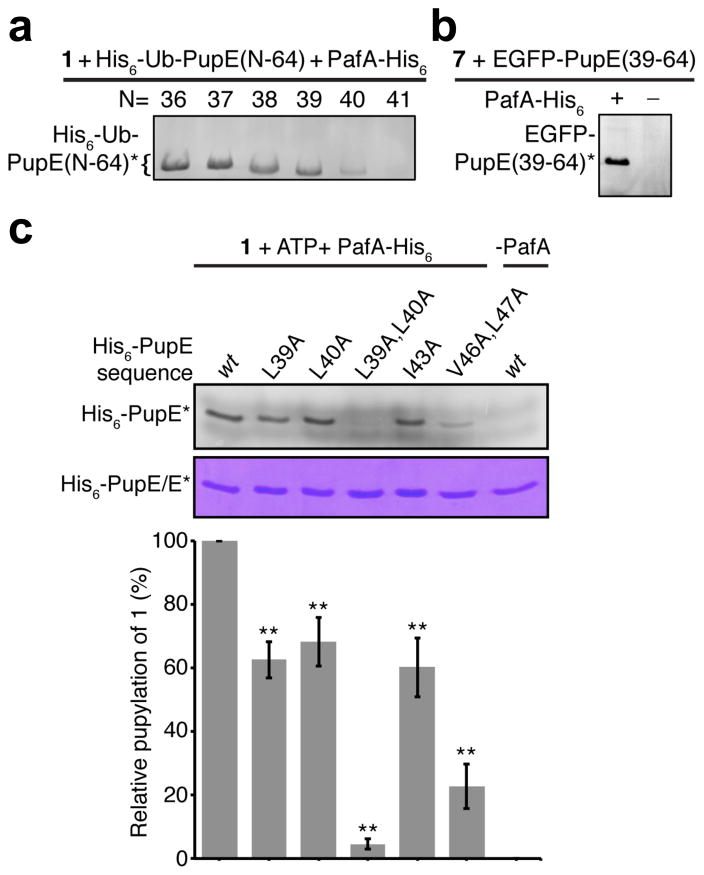
Figure 3. Identification of a minimal sequence and residues in Pup critical for pupylation.
(a) In-gel fluorescence from 15% SDS-PAGE gel showing PafA-His6 catalyzed labeling of the indicated His6-Ub-PupE fragments by probe 1 in cellular lysates. (b) In-gel fluorescence from 15% SDS-PAGE gel showing the PafA-His6 dependent labeling of EGFP-PupE(39–64) by Nα-TAMRA-L-Lys (7) in cellular lysates. (c) 15% SDS-PAGE gel of PafA mediated labeling of wild-type (wt) and mutant full-length His6-PupE polypeptides by probe 1. Top-gel slice shows in-gel fluorescence of labeled proteins and the bottom gel slice shows coomassie staining as a loading control. The bar-graph below shows quantitation of in-gel fluorescence of each mutant relative to wt Pup and is normalized for protein loading. Error bars, s.d. (n= 3), Student’s two-tailed t-test, ** P < 0.05. Asterisks indicate the probe-labeled fluorescent peptides/proteins in each gel.

