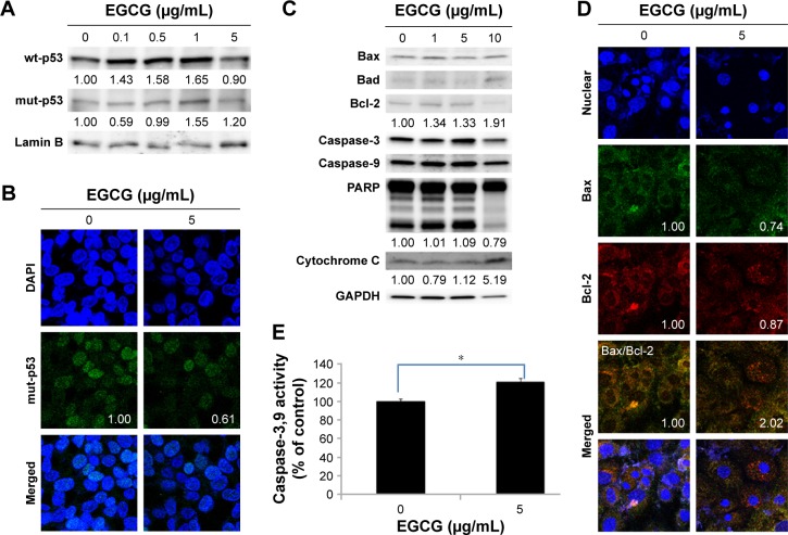
Figure 2.
The apoptosis and necrosis of HuCC-T1 cells by treatment of EGCG.
Notes: Western blot assay: (A) wt-p53, mut-p53; (C) Bax, Bad, Bcl-2, Caspase-3, Caspase-9, and PARP expression. Fluorescence microscopic observation: (B) mut-p53 (Numbers in the boxes indicate intensities of green fluorescence.); (D) Bax, Bcl-2, Caspase-3, Caspase-9, and PARP expression. (E) The extent of Caspase-3,9 activities. Images were observed at 400×. *P<0.01.
Abbreviations: wt, wild-type; mut, mutant; EGCG, epigallocatechin-3-gallate; GAPDH, glyceraldehyde 3-phosphate dehydrogenase; HuCC-T1, human cholangiocellular carcinoma cell line, PARP, poly adenosine diphosphate ribose polymerase.

