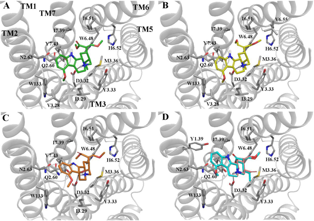
Figure 6.
Docking of (-)-mitragynine and other analogs to the active µ-opioid receptor crystal structure. Top-scoring binding poses of (A) (-)-mitragynine, (B) (Z)-mitragynine, (C) 7-hydroxmitragynine, and (D) antagonists paynantheine (pink) and speciogynine (cyan). Only residue sidechains within 4 Å of the ligand are reported. Polar interactions are shown as dotted lines. TM helices are shown in cartoon representation (in gray). ECL2 and part of TM5 have been omitted for clarity. Residues are labeled using one-letter amino acid code and Ballesteros and Weinstein’s generic numbering scheme.

