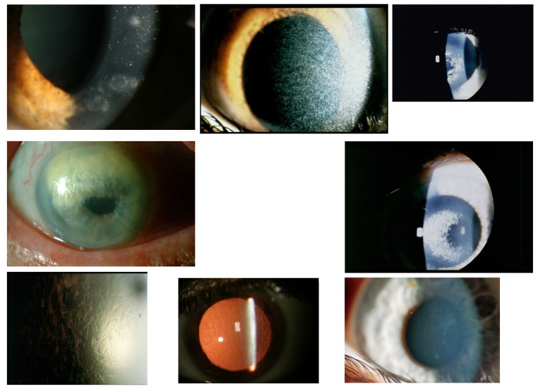
FIGURE 2.
Top row, Case 1 Table 2, MGUS-induced punctiform corneal crystals + patches (left) vs punctiform crystals in cystinosis without patches (middle) and vs crowded comma-shaped crystals of the crystalline type of Schnyder corneal dystrophy + haze (right). Middle row, Case 2 Table 2, MGUS-induced anterior comma-shaped crystals (left) vs crowded comma-shaped crystals of Schnyder corneal dystrophy (right). Bottom row, Case 3 Table 2, MGUS-induced lattice-like lines in retroillumination from the iris (left) vs true lattice lines in retroillumination from the retina of lattice corneal dystrophy type 1 (middle); disappearing of the lattice lines of case 3 and transforming of mild diffuse opacities with focal epithelium elevations after a follow-up of 22 years (right).

