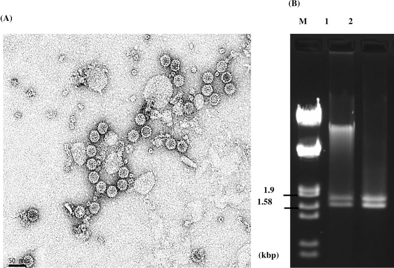
Fig 3. Virus particle morphology and packaged genomic dsRNAs of PdPV-pa.
(A) Particles purified from Pd isolate BB-06, were examined by TEM after negative staining with uranyl formate. The bar indicates 50 nm. (B) Agarose gel electrophoresis profile of PdPV-pa genomic dsRNA segments (lane 1) isolated from the purified virus preparation and the dsRNA segments (lane 2) extracted from mycelia of the same Pd isolate.

