Figure 1.
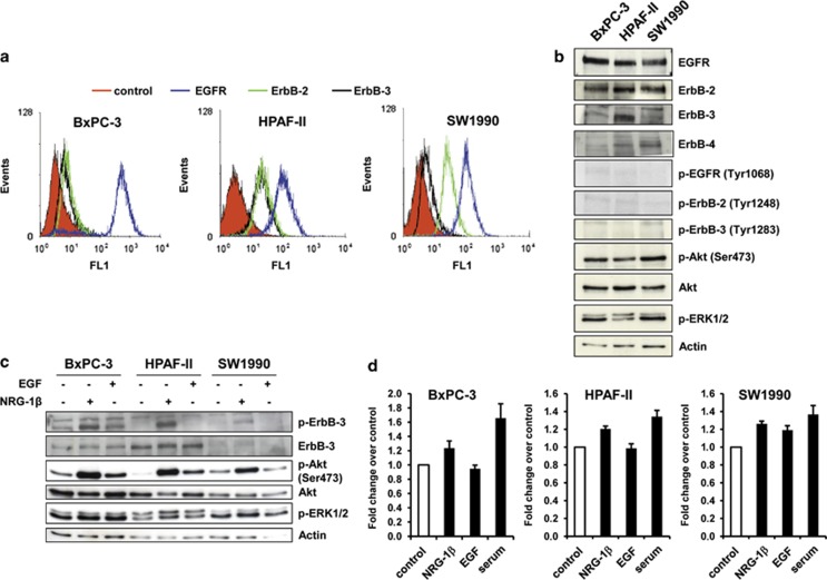
NRG-1β more strongly than EGF activates the ErbB-3/Akt axis. (a) EGFR, ErbB-2 and ErbB-3 surface expression level was analyzed by fluorescence-activated cell sorting analysis. One representative of three independent experiments is shown. (b) Immunoblot analysis of total expression and basal activity of ErbB receptors and downstream signaling pathways. (c) After 24 h of serum starvation, cells were stimulated with either NRG-1β (10 ng/ml) or EGF (20 ng/ml) for 5 min, then cell lysates were blotted as indicated. (d) For cell proliferating assay, 24 h serum-starved cells were grown in the presence or not (control) of NRG-1β (10 ng/ml), EGF (20 ng/ml) or serum (medium containing 10% fetal bovine serum) and stained with MTT after 72 h. Results are expressed as fold change over control.

