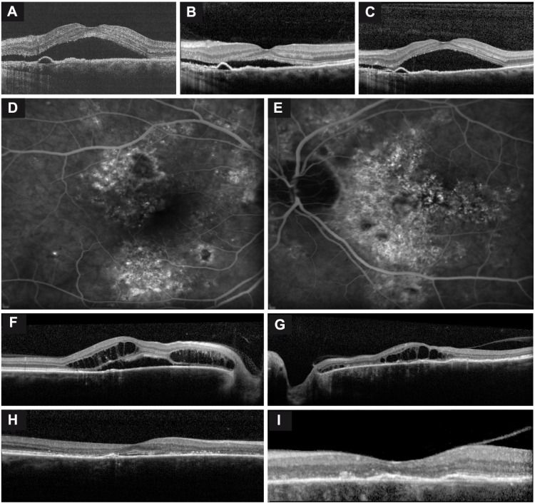
Figure 1.
Imaging of two chronic central serous chorioretinopathy (cCSC) patients.
Notes: Images demonstrate characteristic findings in long-standing cCSC on optical coherence tomography (OCT) and fluorescein angiography (FA). (A–C) Fluctuating subretinal fluid (SRF) accumulation on OCT in the right eye of a patient, and typical subfoveal retinal pigment epithelium detachments. The time between scans A and B was 1 month, in which a clear decrease in SRF occurred, and between scans B and C another 2 weeks elapsed, showing a spontaneous increase in SRF. No therapeutic interventions had been performed between these visits. (D–G) FA and OCT of the right and left eyes of a cCSC patient suffering from bilateral extensive cCSC. On FA, a large area of hyperfluorescence can be seen, indicating advanced disease (D, E). OCT shows not only serous SRF in the right eye but also bilateral central posterior cystoid degeneration, indicative of long-standing disease (F, G). This central posterior cystoid degeneration resolved spontaneously after a period of approximately 6 months (H, I).

