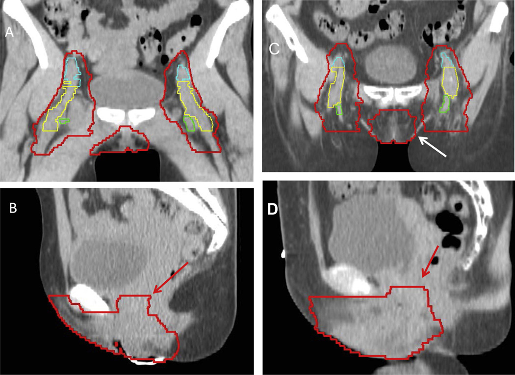
Fig. 3.
A coronal (A) and sagittal (B) slice from a locally advanced case and for the postoperative case (C and D) with the modified consensus contour shown. External iliac vessels are shown in cyan (A), femoral vessels in yellow (A), and saphenous vessels in green (A). Evaluation of coronal and sagittal images is essential for accurate delineation of the vulvar and groin CTV. Coronal images can be useful for identifying the lateral extent of the vulva (white arrow), and on the sagittal images, extension into the vagina is specifically included within the CTV (red arrows).

