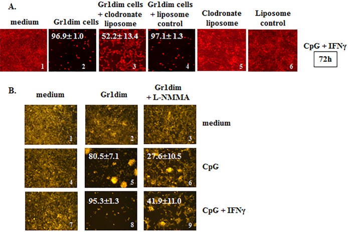
Figure 6. Isolated tumoricidal Gr1dim cells from CY+CpG-treated mice are NO-producing phagocytes.
A. CpG+IFNγ activated myeloid Gr1dim cells from spleens (1×10⁵) of CY+CpG-treated mice were co-cultured with non-irradiated 4T1-f cells (2×10⁴) in the presence or absence of clodronate liposome (25 ng/microwell) or liposomes control at time 0, and tumoricidal activity (% ± SD) was evaluated from 48h to 7 days. Representative photos of the assays at 72h are shown. Data are representative of three technical replicates with similar results. B. Representative photos showing the effect of L-NMMA (iNOS inhibitor, 5mM) upon CpG or CpG+IFNγ activated Gr1dim cells isolated from spleens of CY+CpG-treated mice after 48h in culture. Data are representative of three independent experiments. Images without percentages represent confluent monolayers (killing below detection levels). Images were numerically labeled on the bottom right for the purpose of identification. Photos were taken using an EVOS FL Auto imaging system microscope (A) or a Zeiss Axio Observer A1 microscope (B). Statistical analysis presented in results was performed using Student's t-Test.

