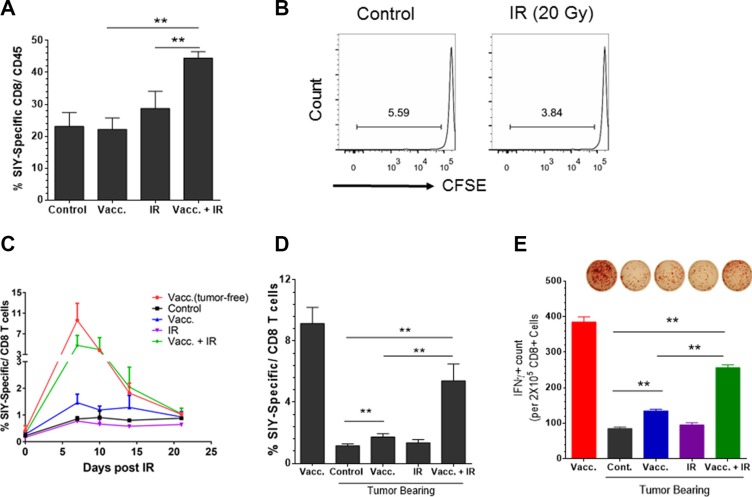
Figure 4. Antigen-specific vaccination plus local radiation convert the established tumor from low level to high level of CD8+ T cell infiltration.
Panc02-SIY fragment tumor-bearing mice were left untreated (control) or received vaccination of 2 × 10⁶ MC57-SIY cells (Vacc.), 20 Gy local ionizing radiation (IR) or vaccination plus IR, respectively. (A) Fragment tumors were harvested and processed for flow cytometric analysis of cell surface markers and SIY-Pentamer staining. Quantitative data of the percentage of SIY-specific T cells among all CD8+ T cells are presented. (B) IR alone fails to prime T cells in DLNs. Panc02-SIY fragment tumor were left untreated or received 20 Gy local IR. CFSE labeled 2C T cells were injected via the tail vein. Five days later, DLNs were analyzed for CD8+ T cell proliferation. (C) Panc02-SIY fragment tumor-bearing mice were left untreated (control) or received the indicated treatments. Peripheral blood (PB) was processed for flow cytometric analysis for CD8 and the SIY-pentamer at the indicated time points. Eight days post treatment, PB, DLNs were used for following studies: (D) PB was evaluated by flow cytometric analysis for CD8 and the SIY-pentamer; (E) CD8+ T cells were isolated from DLNs and incubated with or without the SIY peptide and irradiated naive splenocytes. The culture systems were applied to IFNγ ELISPOT assay. Lymph node cells from at least three mice in each group were pooled together to obtain one sample. Representative images (upper) and Quantitative data (lower) were presented. Error bar are mean ± S.E.M. P values were generated by student's t-test, *P < 0.05, **P < 0.01.

