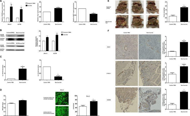
Figure 2. Exosomes derived EOC cells activate macrophages to a TAM-like phenotype in vitro and vivo, which can promote the progression of EOC.
Macrophages were treated with EOC-derived exosomes (100 ug/ml) or control (PBS). (A) After 48 hours, qRT-PCR was applied using primers for CD206, Arg-1, or MCP-1. (B) After 96 hours, the expression of M2 surface marker CD206 was detected by western blot. (C) Cytokine levels in the media of macrophages. (D) Proliferation and migration capacity of Skov-3 human ovarian cancer cells which incubated with the supernatants of macrophages treated with the exosomes or control (PBS) was performed using MTS and Transwell assay. Cell viability was determined by OD measurement and migrated cells were counted. (E) Photographs of appearance of tumor deposits at necropsy of the injected mice. (F) Representative IHC examples of CD31, LYVE-1 and CD206 staining are shown in tumors of Skov3-exo group and compared with control group; scale bar = 50 μm. CD31 positive ‘hotspots’, LYVE-1 positive ‘hotspots’ and CD206-positive cells were calculated as number of positive cells. Data are shown as mean ± SEM, n = 3 independent experiments; *p < 0.05, **p < 0.01, and ***p < 0.001 compared with the control treatments.

