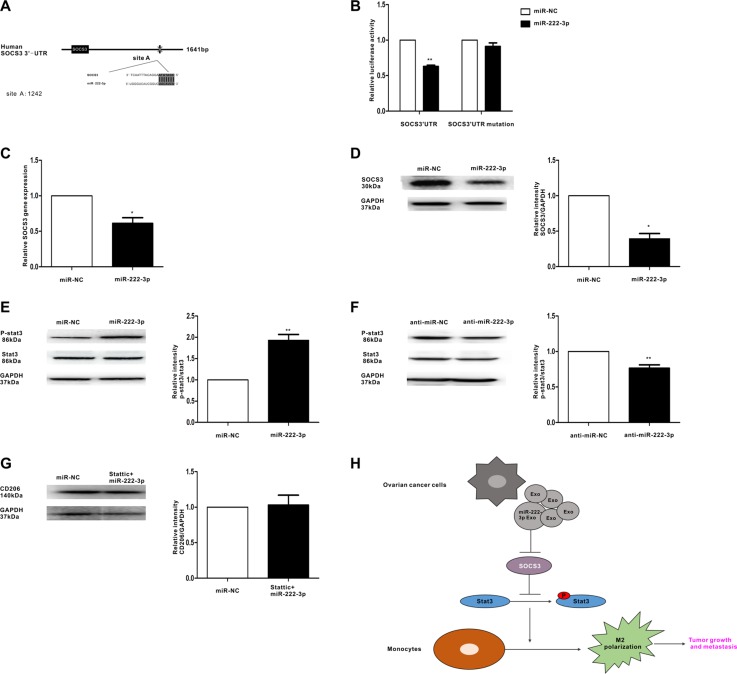
Figure 6. MiR-222-3p downregulates SOCS3 expression and activates STAT3 signaling pathways in macrophages.
(A) Binding sites of miR-222-3p in the 3′UTR of the SOCS3 gene according to bioinformatic analysis. (B) HEK293T cells were co-transfected with the 3′UTR luciferase report plasmid and miR-222-3p mimics or miR-negative control. Then, luciferase activity was measured 40 hours later. Data are presented as relative luciferase activity. (C) Expression of SOCS3 gene in macrophages that were transfected with miR-222-3p or miR-negative control, as performed by qRT-PCR. (D) A representative immunoblot of SOCS3 in macrophages which were transfected with miR-222-3p or miR-negative control. (E, F) The expressions of phosphorylated (p-) STAT3 and total STAT3 in macrophages which were transfected with miR-222-3p mimic and inhibitor were detected by western blot. (G) Macrophages were pretreated with STAT3 inhibitor stattic before transfecting with miR-222-3p mimic or miR-negative control. Expression of M2 marker CD206 was detected by western blot. Data are shown as mean ± SEM, n = 3 independent experiments; *p < 0.05 and **p < 0.01 compared with the miR-NC treatments. (H) Model for the role of miR-222-3p which was enriched in ovarian cancer cells derived-exosomes in the regulation of macrophage polarization in tumor microenvironment.

