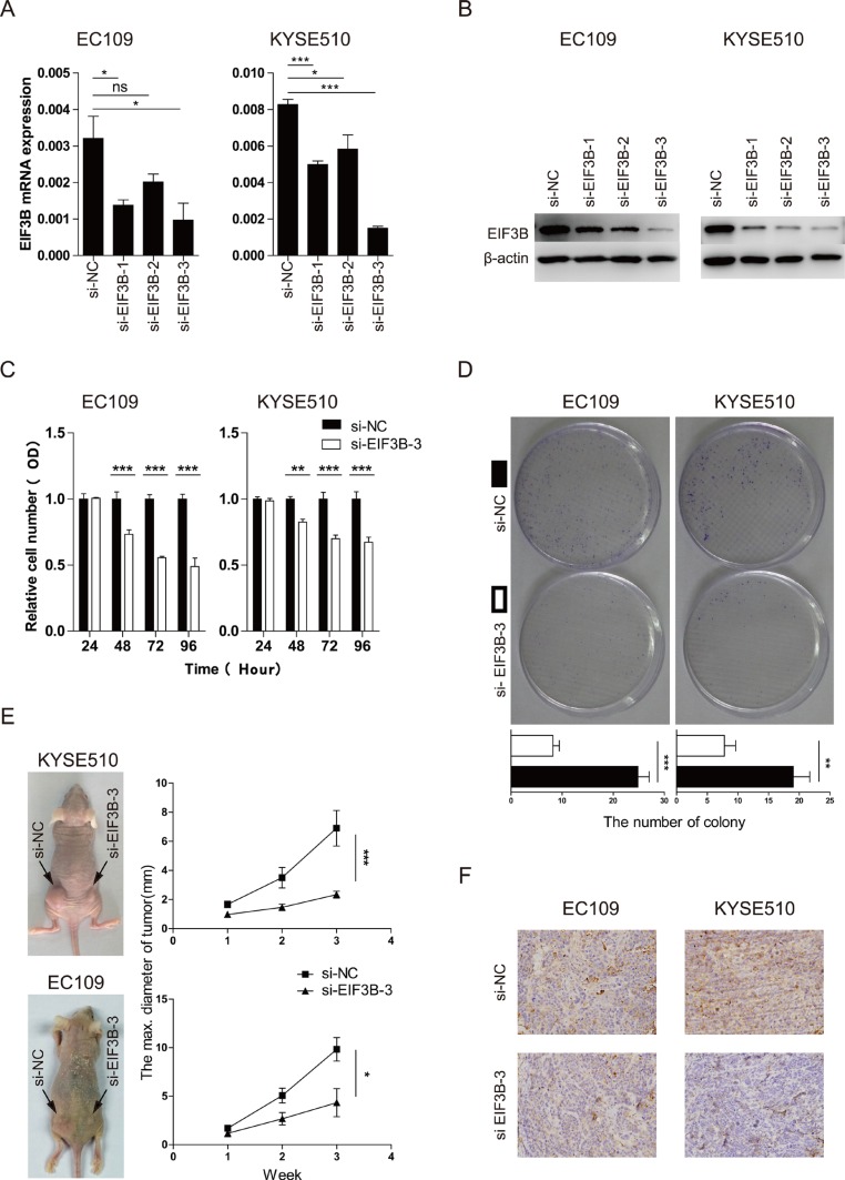
Figure 2. EIF3B promotes the cell proliferation of ESCC.
(A and B), the effect of the knockdown was validated through qRT-PCR and Western blot analyses. β-actin was used as an internal reference. (C), the proliferative ability was assessed with CCK-8 assay at 24, 48, 72, and 96 hours after transfection. (D), the proliferative ability was assessed with colony-forming assay and analyzed statistically after 10-day culture. (E), the proliferative ability was assessed with tumor xenograft assay and analyzed statistically 3 weeks after implantation. (F), the representative staining intensity of EIF3B expression in transplanted tumors was detected with IHC. The values were shown as the mean ± SD. (ns: no significance, *p <0.05, **p <0.01, ***p < 0.001).

