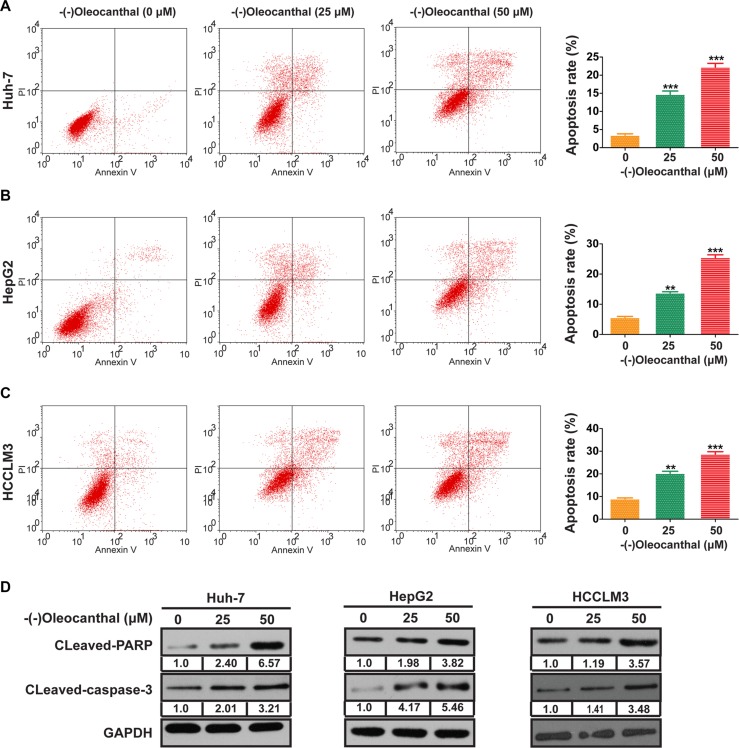
Figure 2. (−)-Oleocanthal induces cell apoptosis in HCC cells in vitro.
(A–C) The representative flow cytometry histograms of cell apoptosis for HCC cells treated for 48 h with increased doses of (−)-oleocanthal. (D) The expression of cleavages of PARP and caspase-3 was explored by western blotting. GAPDH was used as loading control. Data was presented as the means ± SD of three independent experiments. ** compared with control, P < 0.01. *** compared with control, P < 0.001.

