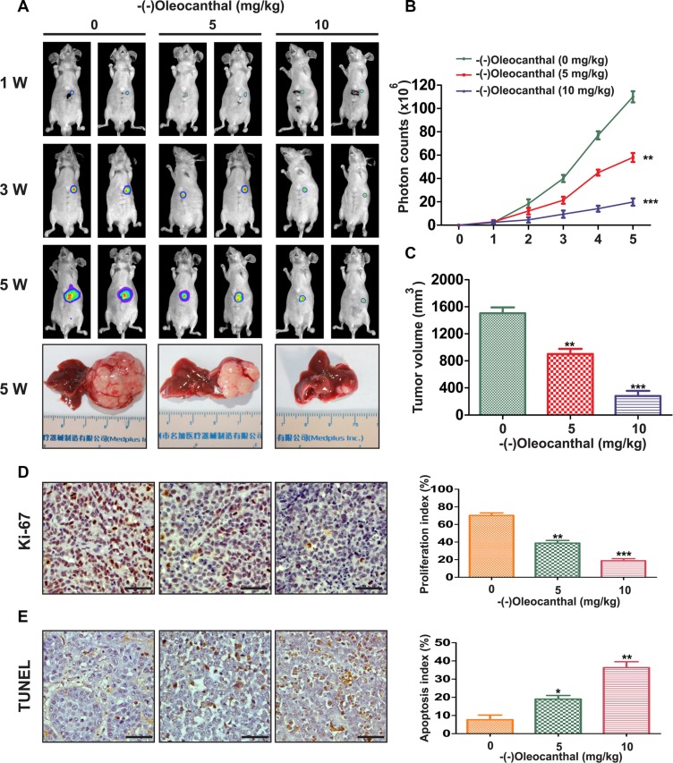
Figure 3. Effects of anti-proliferation and pro-apoptosis by (−)-oleocanthal in an orthotopic tumor model of HCC in vivo.
(A) Representative images of mice from bioluminescent imaging at the first, third and fifth week after (−)-oleocanthal treatment, respectively. The mice were sacrificed at the end of treatment and representative images of gross specimen were shown. (B) Quantification of the tumor growth based on the luciferase intensity. (C) The tumor volumes were measured with vernier calipers. (D) Immunohistochemical analysis of Ki-67 for cell proliferation in tumor tissues. Ki-67-positive cells were counted to evaluate the proliferation index. Scale bars = 200 μm. (E) TUNEL analysis was used to detect the apoptosis in tumor tissues. TUNEL-positive cells were counted to calculate the apoptosis index. Scale bars = 200 μm. Data was presented as the means ± SD of three independent experiments. * compared with control, P < 0.05. ** compared with control, P < 0.01. *** compared with control, P < 0.001.

