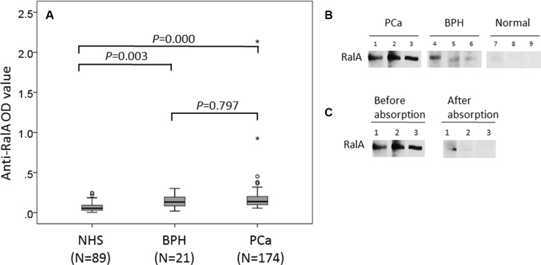
Figure 1. Detection of autoantibodies against RalA in human sera by ELISA and Western blotting analysis.
(A) Autoantibody level to RalA detected by ELISA is expressed as optical density units. (B) Western blotting showed the anti-RalA immunoreactivity of representative sera from three patients with PCa (lanes 1–3), three patients with BPH (lanes 4–6), and three normal human subjects (lanes 7–9). The PCa and BPH sera used for Western blotting were from patients that contain antibodies against RalA as detected by ELISA and the ODvalues in ELISA correlated with the intensity of signals in Western blotting. (C) The serum anti-RalA immunoreactivity of the PCa patients decreased dramatically after pre-absorption with recombinant RalA protein.

