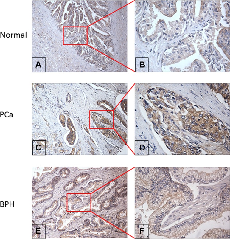
Figure 5. Evaluation of RalA protein expression in normal, PCa and BPH prostate tissue by immunohistochemistry.
(A & B) Moderate positive staining of RalA expression in representative normal prostate tissue at 100× and 400× magnification respectively; (C & D) Weak positive staining of RalA expression in PCa tissue at 100× and 400× magnification respectively; (E & F) Strong positive staining of RalA expression in BPH tissue at 100× and 400× magnification respectively.

