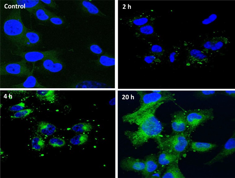
Figure 4. HiMet-C6 EVs are internalized by LoMet-C4 cells.
EV membranes from HiMet-C6 cells were labelled with PKH67 dye as described in “Materials & Methods” and 20 μg/mL of labelled EVs (green) incubated with LoMet-C4 cells (nuclei stained blue with DAPI) for the indicated times. Images are representative of 3 independent experiments. Visualization was performed by confocal microscopy at 63× magnification (Zeiss LSM 510 Meta).

