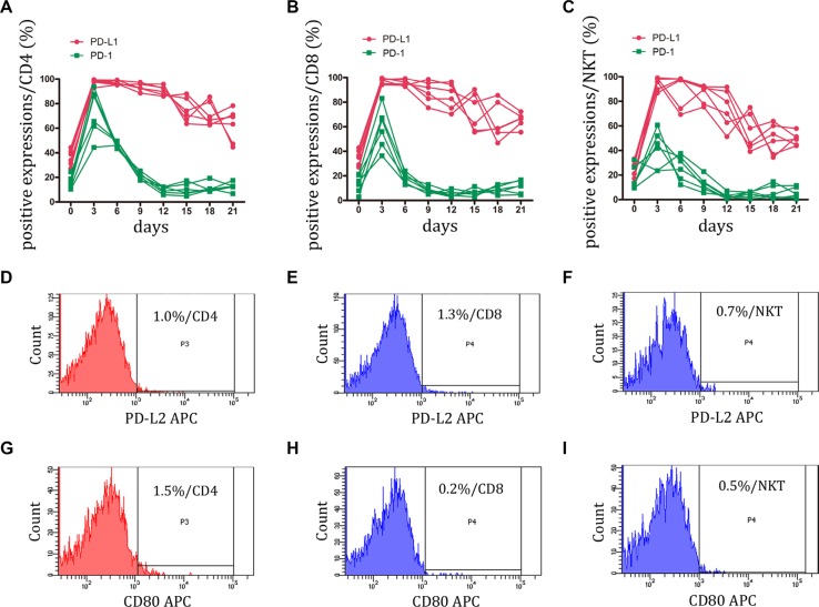
Figure 2. Expression of PD-1 and its ligands, PD-L1 and PD-L2 on CIK cells was detected.
(A–C) During small-scale CIK culture, PD-1 and PD-L1 expressions on CD3+CD4+ T, CD3+CD8+ T and CD3+CD56+ NKT cells were detected by FCM. Data represent six independent experiments. (D–I) PD-L2 and B7-1 (CD80) were almost not expressed by CD3+CD4+ T, CD3+CD8+ T and CD3+CD56+ NKT cells. The representative photos were shown.

