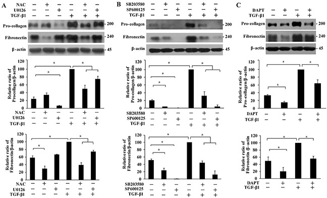
Figure 6. Effect of MAPKs and Notch3 on TGF-β1-induced secretion of extracellular matrix proteins.

IMR-90 cells were pre-treated with NAC (A, 4 mM), U0126 (A, 10 μM), SB203580 (B, 10 μM), SP600125 (B, 20 μM) or DAPT (C, 10 μM) for 1 h, followed by TGF-β1 treatment (200 pM, 48 h). The pro-collagen and fibronectin bands were analyzed by Western analysis, quantified, normalized to those of β-actin, and presented relative to that of TGF-β1 treatment only as means ± SEM (n = 3). *, P < 0.05.
