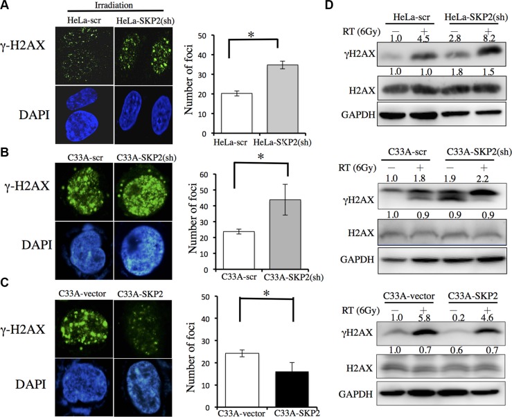
Figure 3. SKP2 increased DNA damages clearance upon ionizing radiation in human cervical cancer cell lines.
C33A-scr, C33A-SKP2(sh), C33A-vector, C33A-SKP2, HeLa-scr and HeLa-SKP2(sh) cells were treated with 6-Gy irradiation. After 4 hours incubated, immunofluorescence staining for γ-H2AX (green) and DNA counterstaining with DAPI (blue). Number of γ-H2AX was counted. Western blots were done to determine the level of γ-H2AX and H2AX. GAPDH was used as the protein-loading control. (A) More γ-H2AX foci were found in HeLa-SKP2(sh) than HeLa-scr cells, after irradiation. The results of western blotting showed greater expression in HeLa-SKP2(sh) than HeLa-scr cells following irradiation. (B) More γ-H2AX foci were found in C33A-SKP2(sh) than C33A-scr cells, after irradiation. The results of western blotting showed greater expression in C33A-SKP2(sh) than C33A-scr cells following irradiation. (C) More γ-H2AX foci were found in C33A-vector than C33A-SKP2 cells, after irradiation. (D) The results of western blotting showed, greater expression of γ-H2AX in HeLa-SKP2(sh), C33A-SKP2(sh) and C33A-vector cells than HeLa-scr, C33A-scr and C33A-SKP2 cells following irradiation, respectively. Lower expression SKP2 of cells associated higher expression of γ-H2AX after irradiation. (*P <0.001).

