
Figure 1. The levels of O-GlcNAcylation, OGT, E6, E7 and O-GlcNAcylation of HCF-1 levels are elevated in cervical cancer tissues.
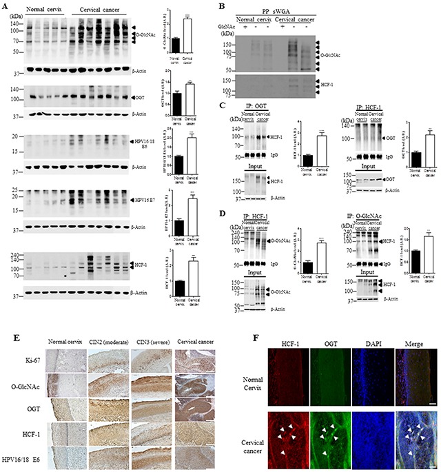
A. Representative Western blot and quantification of O-GlcNAc, OGT, E6, E7 and HCF-1 in normal cervical (n=5) or cervical cancer (n=7) tissues. Band intensity was normalized to β-actin. Data are presented as mean ± SEM. **P<0.05, ***P<0.001 by t test. B. Cell lysates were precipitated using agarose beads coupled to sWGA (PP sWGA) and the precipitates were immunoblotted with an anti-O-GlcNAc – or -HCF-1 antibody. For control, the inhibitory monosaccharide GlcNAc was added during sWGA-lectin-affinity purification. Data are representative of at least 3 independent experiments. Binding of OGT C. or O-GlcNAc D. to HCF-1. Representative immunoblots and quantification of co-immunoprecipitated HCF-1 to OGT or O-GlcNAc in normal cervical or cervical cancer tissues. Tissue lysates were subjected to immuno-precipitation (IP) with an anti-OGT- or - O-GlcNAc antibody and immunoblotted with an anti-HCF-1 antibody. Densitometry of co-immunoprecipitated HCF-1 to OGT or O-GlcNAc was normalized to IgG. Data are presented as mean ± SEM. (n=3 cervical tissues per group). **P < 0.005, ***P < 0.0001 by t test. E. Representative cervical tissue sections stained with an antibody against Ki-67, O-GlcNAc, OGT, HCF-1, E6 or E7 in the normal cervical, CIN2/3 (moderate/severe) and cervical cancer tissues. F. Representative images of double immunofluorescence staining for OGT and HCF-1 plus 4′,6-diamidino-2-phenylindole (DAPI) for nuclear localization. Scale bar, 200 μm.