Figure 5.
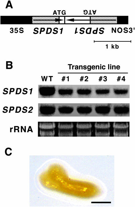
Effect of SPDS1 RNAi in spds2-1. A, Scheme of the SPDS1 RNAi construct. The transcription cassette contains a 0.7-kb promoter region and a part of the N-terminal coding region of SPDS1, the 35S promoter of CaMV, and the nopaline synthase terminator (NOS3′). The hatched boxes represent the promoter region of SPDS1. Arrows indicate the orientation of the SPDS1 gene. B, RNA gel-blot analysis of SPDS1 and SDPS2 transcripts in 10-d-old seedlings of wild-type (WT) and SPDS1 RNAi lines (nos. 1–4). Each lane contained 10 μg of total RNA. rRNA is shown as a loading control. C, Phenotype of the arrested embryo segregated in F2 from the cross between spds2-1 and an SPDS1 RNAi line (no. 1). Bar represents 50 μm.

