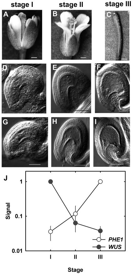
Figure 1.
Developmental stages used for this study. The developmental stages of flowers (A and B) and siliques (C) shown in sections A to C correspond to the pictures of cleared gametophytes and seeds shown below in sections D to I. A, Closed flower (flower was manually opened to show development of gynoecium and anthers). B, Open flower after fertilization. C, Open flower after fertilization. D, Cleared female gametophytes before fusion of central-cell nuclei. E, Cleared seeds containing only endosperm nuclei. F, Cleared seeds containing four-cell embryos. G, Cleared female gametophytes after fusion of central-cell nuclei. H, Cleared seeds containing only endosperm nuclei and one-celled embryo proper. I, Cleared seeds containing eight-cell embryos. Bars: A, 290 μm; B, 360 μm; C, 600 μm; D to I, 50 μm. J, Expression profiles of PHERES and WUSCHEL.

