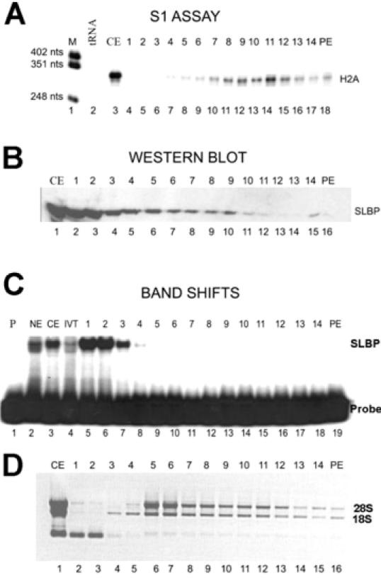
Figure 5.

Association of SLBP with polyribosomes. Cytoplasmic lysate prepared from exponentially growing HeLa cells was fractionated by sucrose gradient centrifugation as described in Materials and Methods. The fraction numbers from the sucrose gradients are indicated above each lane; PE is the sucrose gradient pellet. The monoribosome peak (fractions 6 and 7 in each panel) was identified by ethidium bromide staining of total RNA. (A) Total RNA isolated from each fraction was analyzed for histone H2a mRNA by S1 nuclease mapping. Lane 1, marker (pUC18 digested with HpaII); lane 2, 10 μg of yeast tRNA; lane 3, is analysis of 5 μg of RNA from the cytoplasmic extract; lanes 4–18, sucrose gradient fractions. (B) An aliquot of each fraction sample was analyzed by western blotting for SLBP. Lane 1, 10 μg of cytoplasmic extract; lanes 2–16, sucrose gradient fractions. (C) An aliquot of each fraction was analyzed for RNA binding activity with a mobility shift assay using the radiolabeled stem–loop RNA as a probe. Lane 1, probe; lane 2, nuclear extract; lane 3, cytoplasmic extract; lane 4, in vitro-translated SLBP; Lanes 5–19, sucrose gradient fractions. (D) An aliquot of each fraction was analyzed by agarose gel electrophoresis and the RNAs detected by ethidium bromide staining. Lane 1, RNA from cytoplasmic extract; lanes 2–16: sucrose gradient fractions 1-14 and the pellet (PE).
