
Figure 5.
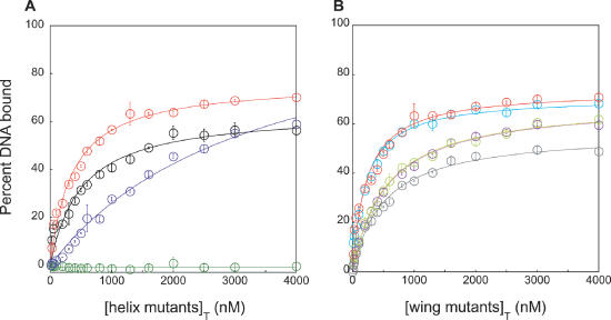
Binding of mthCdc6-1 WHD mutants to the single site. (A) Binding curves for the recognition helix mutants N341A/E342A (black), S330A/S332A (dark blue), R334A/R335A (dark green) and for comparison wild-type protein (red). All curves have been fitted to the independent binding sites model. (B) Binding curves for the wing mutants S355A (light blue), K360A (light green), R362A (purple) and R358A (gray) have also been fitted to the independent binding sites model.