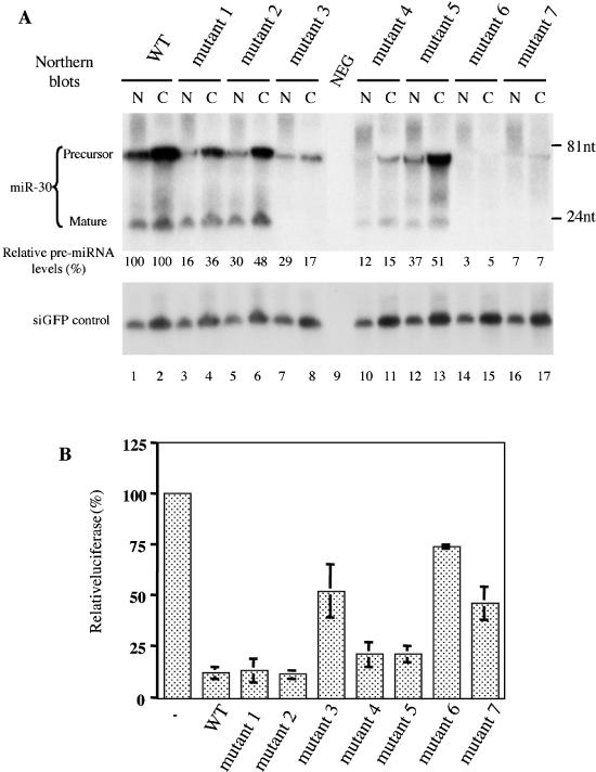
Figure 4.

Expression and function of pre-miR-30 mutants in transfected cells. (A) 293T cells were transfected with 1 μg of pH1-GFP and 1 μg of pSUPER-miR-30 (either WT or one of the mutants) per well in 6-well plates. Two days after transfection, RNAs were isolated from nuclear (N) and cytoplasmic (C) fractions and northern analyses performed. Blots were first probed for miR-30, then stripped and probed for the GFP siRNA. Lane 9: total RNA from untransfected 293T cells (NEG). Intensities of the pre-miRNA bands were quantified by a PhosphorImager, with those of the cytoplasmic and nuclear pre-miRNA expressed from pSUPER-miR-30(WT), both set as 100%. The terminal sequences and predicted secondary structures of the pre-miRNA transcripts are depicted in column 3 of Table 1. Positions of DNA markers are shown at the right side of the autoradiograph. (B) 293T cells in 24-well plates were transfected with 5 ng of the pCMV-Luc-8xmiR-30(P) indicator plasmid, 0.5 ng of the Renilla luciferase internal control plasmid pRL-CMV (Promega) and 20 ng pSUPER or pSUPER-miR-30 (WT or mutants 1 to 7). A dual luciferase assay was performed 2 days after transfection. The firefly luciferase activity (normalized against Renilla luciferase activity) observed upon pSUPER co-transfection was set at 100%. Experiments were performed in triplicate with SD indicated.
