
Figure 3.
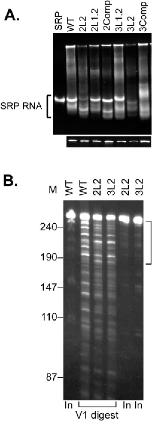
Analysis of wild-type and mutated synthetic SRP RNAs. (A) Native (upper panel) and denaturing (lower panel) 6% PAGE. Equal amounts of RNA were loaded on both gels. The RNAs were visualized by staining with Gelstar®. SRP RNA was extracted from purified canine SRP. The synthetic RNAs are labelled as shown in Table 1. (B) Limited V1 ribonuclease digestion experiments. The digestion products were displayed by 10% denaturing PAGE and the RNA fragments visualized with ethidium bromide staining. The bracket highlights the region that contains RNAs obtained by single cleavages in the Alu portion of SRP RNA. In, 50% of the RNA used in the experiments.