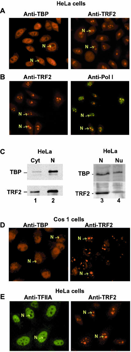
Figure 1.
Distinct localizations of TBP and TRF2 in HeLa cells. (A) HeLa cells were labeled with antibodies against TBP or TRF2 as indicated. (B) HeLa cells were double-labeled with antibodies against TRF2 (red) or pol I (green) as indicated. (C) 20 μg of HeLa cell cytoplasmic (C), nuclear (N), or nucleolar-enriched fraction were separated by SDS-PAGE, and TBP and TRF2 were revealed by immunoblot using the 3G3 and 2A1 antibodies. (D) Cos1 cells labeled with antibodies against TBP or TRF2. (E) HeLa cells were double-labeled with antibodies against TFIIA (red) or TRF2 (green) as indicated. Representative nucleoli are indicated with arrows. Magnification, 40× in all panels.

