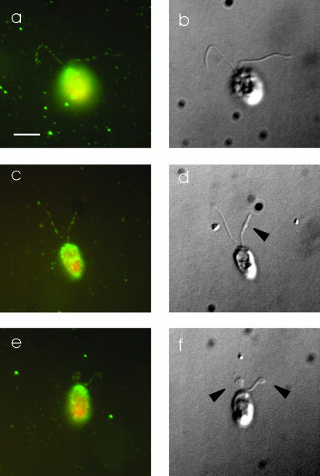
Figure 10.
DHC1b has a normal localization in d1blic mutant cells. Wild-type (a and b) and d1blic mutant cells with near full-length flagella (c and d) or shorter flagella (e and f) were stained with the anti-DHC1b antibody and were imaged by fluorescence microscopy (a, c, and e) or DIC microscopy (b, d, and f). Arrowheads indicate the bulges, shown in Figure 4 to be filled with IFT particles, in d1blic mutant flagella. DHC1b protein is in green and cell bodies in red. The DHC1b protein was present as punctae along the length of both wild-type and d1blic mutant flagella. No obvious accumulation of DHC1b protein was observed in the bulges on the d1blic flagella. Scale bar, 5 μm.

