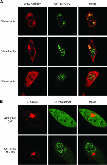
Figure 3.
ERK3 has a nuclear and a Golgi/ERGIC form. (A) HeLa cells were electroporated with the GFP-ERK3 FL vector, plated onto coverslips, and then fixed after 24 h. The slides were stained either with the C-terminal antibody or with the N-terminal ERK3 antibody sc-156. The confocal microscopy images presented are representative of the three patterns observed over the entire field. (B) HeLa cells were electroporated with the GFP-ERK3 ΔFL600 or GFP-ERK3CAT vectors, plated onto coverslips, and then fixed after 24 h. The cells were stained with the ERGIC-53–specific antibody G1/93. The confocal microscopy images are representative cells over the entire field.

