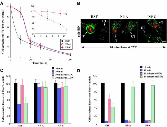
Figure 3.
Recycling of Tfn in normal HSFs versus NPFs. (A) Cells were labeled with 125I-Tfn for 1 h at 16°C and acid-stripped, and the amount of cell-associated Tfn was quantified after various times at 37°C. (B–D) Cells were cotransfected with the DsRed2-Nuc plasmid and either DN rab4 or DN rab11 constructs. After 48 h, the cells were labeled for 1 h at 16°C with Alexa 488-Tfn, washed, acid-stripped, and then incubated for 0, 10, or 40 min at 37°C in the presence of excess unlabeled holo-Tfn (1 mg/ml). The amount of cell-associated Alexa 488-Tfn that remained cell-associated after 10 min (C) and 40 min (D) was quantified by image analysis and expressed as a percentage of the initial fluorescence at 0 min. UT, untransfected; T, transfected. Bar, 20 μm. Values are mean ± SD (n ≥ 60 cells; three independent experiments).

