Figure 3.
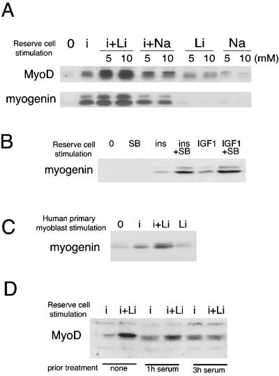
Insulin and lithium chloride (or SB216763) cooperate to activate and induce the differentiation of quiescent reserve cells. (A) C2.7 reserve cell stimulation: C2.7 myoblasts were cultured in 15% serum proliferation medium for 3 d, and then in 3% serum differentiation medium for 4 d. At that time, the supernatant from these cells was diluted with the same volume of serum-free DMEM and after 2 d, the supernatant was again diluted twice with serum-free DMEM. After a total of 8 d of differentiation, undifferentiated quiescent reserve cells were isolated after removal of the myotubes by limited trypsination. The residual adherent reserve cells were incubated in DMEM for 4 h to allow respreading. Shown are Western blots for MyoD and myogenin after 24-h stimulation of reserve cells by different treatments, as indicated: no treatment (0), or insulin alone at a concentration of 3 μg ml-1 (i), LiCl alone at 5 or 10 mM (Li), or insulin and LiCl 10 mM (i+Li). NaCl (Na or i+Na) was used as a control for LiCl addition. (B) Mouse C2.7 reserve cells were treated as described in A. SB216763 was used instead of LiCl at a concentration of 3 μM (SB). Also shown is the effect of 10 nM IGF-1 instead of insulin. (C) Human primary reserve cell stimulation: human reserve cells purified as described in Materials and Methods were treated with insulin and/or LiCl for 24 h in serum-free DMEM before analysis for myogenin expression. Shown is a representative result repeated in three independent experiments. (D) Mouse C2.7 reserve cells were isolated as for Figure 3A, cultivated in DMEM for 4 h to respread on the dish, and then stimulated with serum, at a final concentration of 15% for the indicated times, to reenter the cell cycle, before 24-h stimulation with insulin alone at 3 μg ml-1 (i) or insulin and LiCl at 10 mM (i+Li). Cells were harvested and analyzed by Western blot for MyoD expression.

