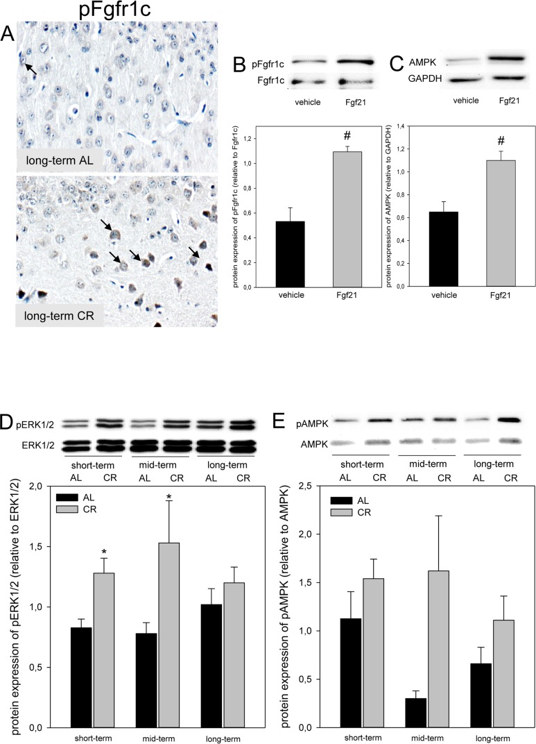
Figure 3.
(A) Representative immunohistochemical images (original magnification x400) of pFgfr1c expression in brain of long-term ad libitum- (AL, upper panel, indicated by arrows) and of caloric-restricted-fed (CR, lower panel, indicated by arrows) ApoE−/− mice. Representative Western blots as well as densitometric analysis of (B) pFgfr1c and (C) AMPK expression in primary glial cells, which were treated with vehicle (DMEM/F12) and 5 μg/ml Fgf21. Representative Western blot and densitometric analysis of (D) pERK1/2 and (E) pAMPK expression in brain of ApoE−/− mice. Mice were fed either AL or CR (60% of ad libitum) for a short-term (4 weeks; n=14), mid-term (20 weeks; n=14) or long-term (64 weeks; n=14). Signals were corrected to that of either ERK1/2 or AMPK. Values are given as means ± SEM; ANOVA, post-hoc pairwise comparison tests: * p < 0.05 vs. AL, # p < 0.05 vs. vehicle.

