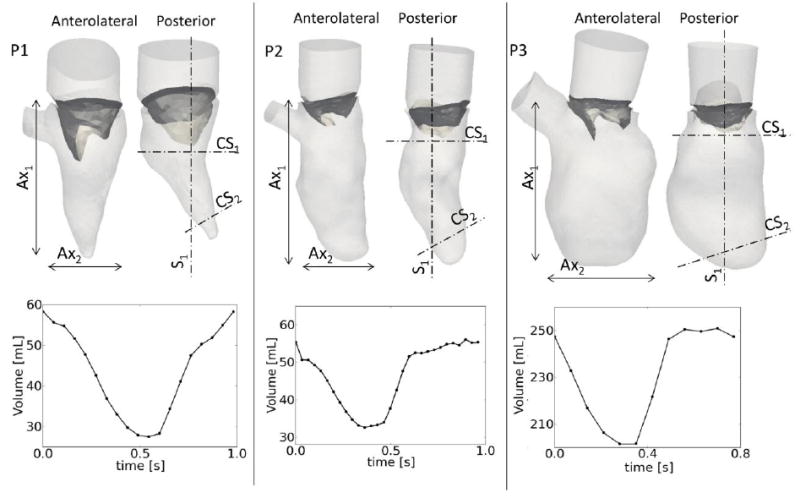
Figure 1.

upper panel: patient geometries, showing the anterolateral and posterior walls of the LV. The dimensions of the major (Ax1) and minor (Ax2) axes are reported in table 1. Lower panel: LV volume curves calculated from the segmented ventricles. Sections S1, CS1 and CS2 used to report relevant results in figure 2 and 3.
