
Figure 1.
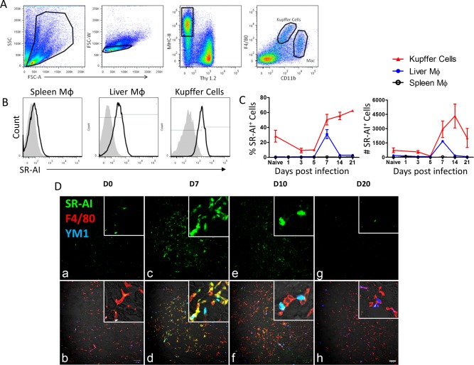
SR‐AI is up‐regulated on Mφ following hepatic viral infection. (A) Flow cytometry gating strategy for liver macrophages. Mononuclear cells were separated from whole‐liver homogenate by density gradient centrifugation, and live singlets were gated on Thy1.2–MHC‐II+. F4/80hiCD11bmid cells were identified as liver‐resident Kupffer cells and F4/80midCD11bhi cells were identified as nonresident macrophages. (B) SR‐AI surface expression (black trace) versus isotype control (gray histogram) in spleen Mφ, liver Mφ, and KCs at day 7 postinfection. (C) Time course of frequency and number of SR‐AI+ cells (determined by gating on isotype control) during AdOVA infection. Data points are mean ± SEM of n=3 mice. (D) Immunofluorescence microscopy of sections from AdLacZ infected mouse liver at 0, 7, 10, and 20 days postinfection (100× magnification and scale bar = 100 μm; insert, ×200 magnification). Panels (A), (C), (E), and (G) show SR‐AI single‐surface staining in green; panels (B), (D), (F), and (H) show merged staining of SR‐AI (green), F4/80 (red), and YM1 (blue).