Figure 3.
Identification of the Microtubule Binding Domain of AtMAP65-1.
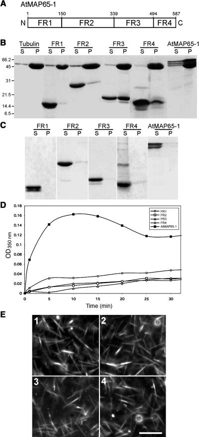
(A) Diagram showing the positions of the four AtMAP65-1 fragments (FR1, FR2, FR3, and FR4). The numbers indicate the positions of the first and the last amino acids of each fragment in the AtMAP65-1 full-length sequence. N and C indicate the N and C termini, respectively.
(B) Cosedimentation of AtMAP65-1 and fragments 1 to 4 with taxol-stabilized microtubules. Microtubules on their own or as a mixture with AtMAP65-1 or AtMAP65-1 fragments 1 to 4 were centrifuged at 100,000g and then the supernatants (S) and pellets (P) were separated on an SDS-PAGE gel and stained with Coomassie.
(C) Sedimentation of the same AtMAP65-1 and AtMAP65-1 fragments 1 to 4 (as in [B]) in the absence of microtubules. AtMAP65-1 and fragments 1 to 4 were analyzed as in (B).
(D) Effect of AtMAP65-1 and fragments 1 to 4 (each 20 μM) on the turbidity of a 20-μM tubulin solution. The assay was performed at 32°C, and the turbidity was monitored at 350 nm.
(E) Dark-field microsopy images of a 20-μM tubulin solution polymerized in the presence of 10 μM AtMAP65-1 fragments 1 to 4 (panels 1 to 4, respectively). Bar = 1 μm.

