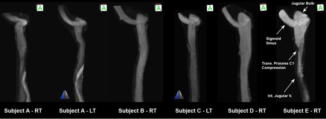
Figure 4.
Anterior view of internal jugular veins of five subjects identified by letters from A to E from combination of right (RT) and left (LT) IJVs sorted from left to right with increasing elevation of jugular bulb from the flooring of the sigmoid sinus junction. The two IJVs with the mirror sign next to them are left-sided IJVs that are mirrored and shown as right-sided IJVs for comparing convenience.

