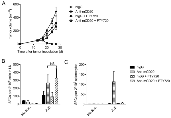
Figure 4. Tumor-specific T cells are primed in the draining lymph node.
(A) A20-bearing WT BALB/c mice (n = 5/group) were administered 100 μg of anti-mCD20 or hIgG on day 11. FTY720 was administered every two days starting on day 10 through the end of the experiment. (B-C) Eleven days after the Ab treatment, DLN cells (B) or splenocytes (C) were collected, and the IFN-γ ELISPOT assay was performed with medium or A20 cell restimulation. The mean ± SEM values are shown. Two-tailed Student’s t test was used for statistical analysis.

