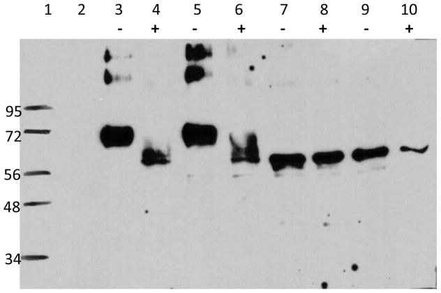
Fig. 1. Evidence for Pfs48/45 N-linked glycosylation.
Mammalian (HEK293T) cells were transfected with Pfs48/45 DNA plasmids and cultured in the presence or absence of tunicamycin for 48 hours. Cell lysates were analyzed by SDS-PAGE under reducing conditions. Lane 1 shows molecular weight standards, lane 2 shows results with un-transfected cells (no DNA). Results with cells transfected with WT (lanes 3, 4), SYN (lanes 5, 6), MUT1 DNA (lanes 7, 8) and MUT2 DNA (lanes 9, 10) are shown in cells cultured in the absence (-) and presence of tunicamycin (+). Equal number of cells lysed in SDS-PAGE sample buffer were loaded in each lane.

