. 2016 Dec 20;46(4):291–296. doi: 10.5624/isd.2016.46.4.291

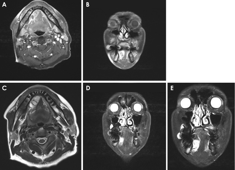
Fig. 4. A. The mass shows heterogeneous intensity with peripheral enhancement in gadolinium-enhanced T1-weighted magnetic resonance imaging (axial view). B. The mass is located superior to the mylohyoid muscle in gadolinium-enhanced T1-weighted magnetic resonance imaging (coronal view). C. Well-defined multilocular cystic mass in the right sublingual space is demonstrated in fat-suppressed T2-weighted magnetic resonance imaging (axial view). D. The mass is surrounded by a hypointense signal, which was suspected to represent a fibrous capsule, in fat-suppressed T2-weighted magnetic resonance imaging (coronal view). E. The location and size of the mass is clearly shown in fat-suppressed T2-weighted magnetic resonance imaging (coronal view).

Fig. 4