FIG 2.
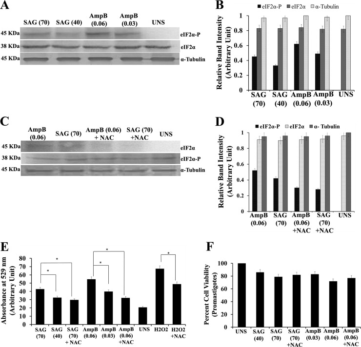
SAG and amphotericin B also induce LdeIF2α phosphorylation. (A) Western blotting to determine the phosphorylation status of LdeIF2α upon exposure to the antileishmanial drugs sodium antimony glucamate (SAG) and amphotericin B (Amp B). L. donovani promastigotes were exposed two different doses, viz., SAG40 and SAG70 for SAG and Amp B0.06 and Amp B0.03 for Amp B for 8 h and immunoreacted with rabbit polyclonal anti-eIF2α (pS51)-phosphospecific antibody. Parasites without any stress served as controls (UNS). Protein loading was monitored by using an anti-Leishmania-specific eIF2α antibody and an anti-α-tubulin antibody. (B) Relative phosphorylation status determined by densitometry. (C) Western blotting to determine LdeIF2α phosphorylation upon exposure to SAG70, SAG plus NAC, Amp B0.06, or Amp B plus NAC after 8 h of treatment. (D) Densitometric analysis of band intensities. (E) ROS generation determined spectrofluorimetrically by using H2DCFDA dye under the above-mentioned conditions. L. donovani promastigotes exposed to H2O2 (200 μM) served as positive controls for ROS measurement. (F) Percent viability of promastigotes exposed to SAG40, SAG70, SAG70 plus NAC, Amp B0.03, Amp B0.06, or Amp B0.06 plus NAC. UNS parasites were used as controls. Values are the means and SDs of results from three independent experiments. (Significant differences are indicated by * [P < 0.05].)

