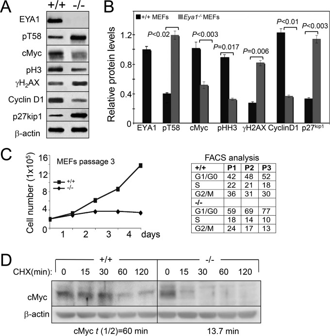
FIG 9.
EYA1 regulates Myc stability in MEFs. (A) Western blot of cell lysates from wild-type (+/+) and Eya1−/− MEFs at passage 2. (B) Quantification of Western blot in panel A. The error bars represent SD, and the P values were calculated using StatView t tests. (C) (Left) Graphic representation of wild-type and Eya1−/− MEFs from passage 3 on the indicated days. Eya1−/− MEFs stopping growing at passage 3; 2 × 10⁵ cells were plated in a 60-mm plate (in triplicate) and were counted after each day of growth for 4 days. The numbers were consistent in triplicate, and averages are shown. (Right) Flow cytometry analysis of wild-type and Eya1−/− MEFs at passages 1, 2, and 3. (D) Western blot analysis of cell lysates from passage 2 wild-type or Eya1−/− MEFs collected at the indicated time points after treatment with CHX. The cells were starved in low serum for 3 days before treatment with CHX. t(1/2), half-life of cMyc.

