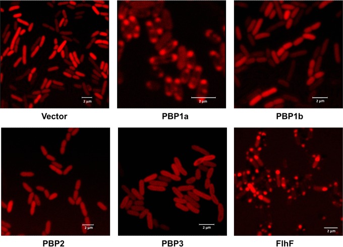
FIG 5.
Subcellular location of HMM PBPs in PA14. P. aeruginosa PBPs were fused with mCherry at the N terminus of each protein and transformed into PA14. One microliter of an overnight culture was spotted on a slide and covered by a thin layer of 1.2% agarose. A known polar protein, FlhF, was used as a polar control. The empty vector expressing mCherry was used as a negative control. The location of fusion proteins was observed using a Leica SP2 confocal microscope.

